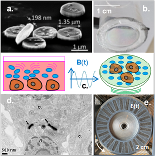
a. dispersed NiFe particles (SEM image) b. PDMS membrane with embedded array of MPs. c. sketches of pancreas cells stimulated by i) dispersed MPs, ii) MPs embedded in PDMS membrane. d. TEM image of INS-1E cells + MPs, internalized after an applied field B(t) of 40 Hz (SYMMES). f. Rotating magnetic field source: 24 NdFeB magnets in a circular rotating plate.
In the context of intensive research on treatments against diabetes – major health threat to millions of people, we report here on an innovative approach based on mechanobiology. Basically, chronic hyperglycemia is regulated by a supply of insulin, if insufficiently secreted. We show here that the mechanical stimulation of pancreatic beta cells, via the vibration of magnetic microparticles – either dispersed among the cells or embedded in a polymer membrane -, can trigger the insulin secretion.
Firstly studied for the destruction of cancer cells (Kim et al, Nat. mat., 9, 2010; Leulmi et al, Nanoscale, 7, 2015), our magnetic particles (MPs) composed of gold-coated permalloy (Ni80Fe20) disks, in a vortex magnetic state, have been tested here for stimulating the insulin production function of pancreatic beta cells. Magnetically actuated by a rotating magnetic field of low frequency, their vibrations exerted mechanical forces on the beta cells (INS-1E β-islet pancreatic cells). The magnetic disks – Au/NiFe/Au – of 1.3 µm diameter, with NiFe thicknesses of 60 to 200 nm, were first used dispersed in the cell culture medium, deposited on the surface of the pancreatic cells, and submitted to the rotating magnetic field.
The cell response to this mechanical stimulation shows an increase in the amount of insulin produced, versus the basal insulin secretion from unstimulated cells. The insulin secretion depends on the concentration of particles, frequency of the magnetic field and duration of the stimulation. A significant increase in insulin release was observed in particular for the largest particles concentration (50 µg.mL−1) and field frequencies (20 to 40 Hz), with exposure times between 10 and 30 min. Interestingly, this magnetomechanical actuation potentially triggered the internalization of the particles into the cell cytoplasm, as observed by TEM (Transmission electron microscopy) imaging.
A second type of magnetic actuation was investigated, less invasive, consisting of a global mechanical stimulation of the pancreatic cells, grown on a flexible polymer magnetoelastic membrane. In this second approach, similar magnetic particles were embedded in a 5 µm thick PDMS (polydimethylsiloxane) membrane. The rotating magnetic field induced the membrane vibration, globally transmitted to the pancreatic cells. In this configuration, the insulin secretion was again significantly enhanced by the mechanical stimuli.
In both cases, the amount of insulin secreted can be comparable to an effective glucose response. We also controlled that this magnetomechanical treatment did not lead to the pancreatic cell death. While some diabetes still require invasive treatments based on daily insulin subcutaneous injections, this mechanotransduction effect, remotely triggered, may open new doors towards diabetes treatments.
Teams: HEALTH AND BIOLOGY
Collaboration: IRIG/SYMMES, Y. Hou (biochemistry); M. Carriere (biology) marie.carriere@cea.fr
Funding: EU’s H2020 Fet Open ABIOMATER project No 665440
Further reading: Magnetic particles for triggering insulin release in INS-1E cells subjected to a rotating magnetic field,S. Ponomareva, H. Joisten, T. François, C. Naud, R. Morel, Y. Hou, T. Myers, I. Joumard, B. Dieny, M. Carriere, Nanoscale, 14, 13274 (2022). Open access: hal-03841954.
Contact: Helene Joisten, Bernard Dieny
